
Evidence
Last reviewed 6 September 2026 · Trust Center / change history
A claim is only as strong as its study design.
September intelligence integrated: VENT-SIM, CO₂ Modulation in EVT and CHILL-AP are now reflected in the research register and clinical-development planning.
AIRCHILL follows a staged evidence ladder: measured mechanism and preclinical safety define a strong rationale, and each next study is designed to turn that rationale into reproducible product performance and patient-relevant evidence. Evidence levels remain explicit so a promising signal is never confused with a result that has already been clinically validated.
Research corpus · frozen 10 August 2026
33,808 records — but evidence strength is not a record count.
Our hypothermia research corpus contains 32,462 deduplicated PubMed records and 1,331 unique ClinicalTrials.gov studies, plus project sources, guidelines and targeted registry checks. Only 60 sources are currently manually included and documented for result claims. Protocols, primary reports, follow-ups and secondary analyses can belong to the same study family, so they are linked rather than counted as independent evidence.
Two studies that both use 33 °C can test different interventions if one starts earlier, reaches target faster, treats longer, cools another compartment or rewarm differently. Population, comparator and co-interventions such as sedation, shivering control and fever prevention are part of the interpretation.
Research-method rule: database records are not independent study populations. Register entries describe planning and status, not clinical benefit. Negative and neutral studies remain visible.
The rationale
Why anyone cools a brain at all.
The first three links of this chain are physiology and are not in dispute. The fourth is the one the whole field has been trying to close for twenty years, and it is the reason this company exists. We draw it differently on purpose.
The 6–7 % per degree figure is the standard physiological estimate for cerebral metabolic rate, quoted here as mechanism and not as a clinical result. Everything downstream of it — whether a measurable temperature drop translates into a measurable patient benefit — is the open question — and it is the question this company was built to answer. We publish the evidence that points our way and the evidence that does not, and we do not treat either as settled.
Why we expect this to work
Three lines of evidence, one direction.
The animal models, the human timing signal and our own device data all point the same way. Below them stands what would argue against us, because a case that hides its counter-examples is not a case.
Swipe sideways to see all three →
Clinical context
TTM2 reached target temperature about five hours in. It cannot answer the early question.
TTM2 randomised patients a median of 136 minutes after return of circulation and then needed a median of three more hours to reach 34 °C. What it compared was late temperature control against fever control, and it found no mortality benefit while reporting more arrhythmias with haemodynamic compromise. No arm of that trial cooled within the first minutes on scene. A neutral result for a late intervention is not a result for an early one — the early question was never in the design.
136 minutes, then three hours
Median time from return of circulation to randomisation 136 minutes; median time from randomisation to 34 °C three hours, interquartile range two to four. Target temperature therefore around five hours after the event.
Late cooling is answered
Routine cooling to 33 °C begun hours after the event does not improve outcomes. We state that plainly and we do not build anything on it.
The first-minute effect remains a distinct clinical-validation target
TTM2 says nothing about a temperature effect produced in the first minutes, because it never produced one. Using it either for or against ultra-early cooling is a misreading of the trial.
This is the counterweight, and we state it before anyone else does. PRINCESS cooled transnasally during resuscitation in 677 patients across 11 EMS systems in seven countries and missed its primary endpoint. Taccone et al., Crit Care 2024;28:335 filtered the HACA-like shockable subgroup out of TTM2 (n = 600) and found no advantage there either — which hits our own target-group logic directly. So ultra-early selective cooling remains a live, testable clinical hypothesis. The next step is not to repeat earlier trials unchanged, but to test a defined phenotype, a distinct thermal exposure and a prospectively powered endpoint that can measure and size the effect.
Dankiewicz J, Cronberg T, Lilja G et al. Hypothermia versus Normothermia after Out-of-Hospital Cardiac Arrest. N Engl J Med 2021;384:2283–2294 (TTM2, n = 1,861; median time to randomisation 136 minutes in the hypothermia arm and 133 in the normothermia arm, measured from sustained return of circulation; median three hours from randomisation to 34 °C, IQR 2–4) · Nordberg P, Taccone FS, Truhlář A et al. JAMA 2019;321:1677–1685 (PRINCESS) · Taccone FS, Cariou A, Zorzi S et al. Crit Care 2024;28:335 · ERC-ESICM Post-Resuscitation Care Guidelines 2025.
The pattern
Every estimate the field has produced, with the clock on it.
One axis, nine estimates, and on every row the time from the event to the start of cooling. That column is the argument.
Swipe the chart sideways to see the intervals →
The case for cooling
The other half of the literature, with its own footnotes.
A site that only quoted the neutral trials would be as one-sided as one that only quoted the positive ones. Cooling does have evidence in its favour. It is thinner than its advocates say and stronger than its critics admit — and every figure below carries the caveat that comes with it.
HYPERION · NEJM 2019
Non-shockable rhythm, n = 584. Good neurological outcome at 90 days 10.2 % with 33 °C against 5.7 % with normothermia — an absolute difference of 4.5 percentage points (95 % CI 0.1–8.9, p = 0.04). Fragility index 1: one patient changing outcome would have removed the significance. No mortality benefit (81.3 % vs 83.2 %).
Cochrane · 2023
Conventional cooling to 32–34 °C against standard care: RR 1.60 for good neurological outcome (95 % CI 1.15–2.23), 8 trials, 2,870 participants. GRADE certainty: low. No effect on survival, and more pneumonia, hypokalaemia and severe arrhythmia in the cooled arm.
ERC-ESICM · 2025
Temperature control stays in the guideline: active fever prevention at or below 37.5 °C, 36–72 hours of control, and explicitly no routine prehospital cold fluid infusion. The debate is about the target and the timing, not about whether temperature matters.
The largest dataset on the question
And then 33,933 patients from the German Resuscitation Registry.
Knapp et al. analysed every adult in the registry between 2006 and 2022 who was comatose on admission after cardiac arrest — a catchment of more than 31 million people in Germany and Austria. 10,034 received mild therapeutic hypothermia, 23,899 did not. A multivariable logistic regression adjusted for every known outcome variable, including witnessed arrest, CPR duration, coronary angiography and hospital caseload.
HYPERION shows a benefit that hangs on a single patient. Cochrane shows an effect with low certainty and no survival gain. The registry shows an association across 33,933 patients but, in the authors’ own words, “can only reveal associations; the ultimate cause of the effect remains unknown” — patients who get cooled are also the patients who reach a cardiac arrest centre. TTM2 shows no benefit, but only reached target temperature about five hours in. Four results, four different weaknesses, no settled answer. And not one of them compares early cooling against late cooling: they compare cooling against no cooling. The timing question is simply unanswered — which is why it is ours. Our own claim register keeps ultra-early selective cooling at clinical hypothesis, and it will stay there until a patient study says otherwise.
PRINCESS2
About 1,022 patients. Selective on-scene cooling in shockable rhythm. The 100-patient pilot reported 92 % protocol adherence and no serious device-related events.
ICECAP
1,158 patients in an adaptive randomized comparison of cooling duration at 33 °C. Longer cooling up to 72 hours did not improve neurological outcome. Because every arm received 33 °C hypothermia, ICECAP answers a duration question — not hypothermia versus normothermia.
OverCool
Feasibility work on ultra-fast cooling through the respiratory route, including liquid ventilation. The closest active programme to our own question.
COTTIS 2
Transnasal cooling in stroke and thrombectomy — the same device family as RhinoChill, a different indication.
Lascarrou JB, Merdji H, Le Gouge A et al. Targeted Temperature Management for Cardiac Arrest with Nonshockable Rhythm. N Engl J Med 2019;381:2327–2337, doi 10.1056/NEJMoa1906661 · Arrich J, Schmutz R, Warenits AM et al. Hypothermia for neuroprotection in adults after cardiac arrest. Cochrane Database of Systematic Reviews 2023, CD004128.pub5 · ERC-ESICM Post-Resuscitation Care Guidelines 2025 · PRINCESS2, NCT06025123; Dillenbeck E et al., Crit Care 2026 (pilot) · ICECAP, NCT04217551 · OverCool, NCT06798818 · COTTIS 2 · Knapp J, Steffen R, Huber M, Heilman S, Rauch S, Bernhard M, Fischer M. Mild therapeutic hypothermia after cardiac arrest — effect on survival with good neurological outcome outside of randomised controlled trials: a registry-based analysis. Eur J Anaesthesiol 2024;41(10):779–786, doi 10.1097/EJA.0000000000002016 (open access). The registry study is observational: patients selected for cooling may differ systematically from those who were not, and treatment in a university hospital or cardiac arrest centre could only be adjusted for indirectly. It also says nothing about when cooling was started.
The map
Where cooling has been tried, and where it has not.
Eight indications for which the start time is documented, plotted against the evidence status. The left-hand column is where our trial goes.
Swipe the map sideways →
Our own measurements
The effect follows the airway.
This is the figure that sets the boundary of what we may claim today, and the reason the first product targets selective cooling rather than a whole-body claim.
Swipe the chart sideways →
The measurement itself
Two methods, one direction.
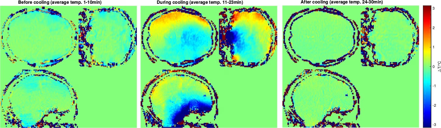
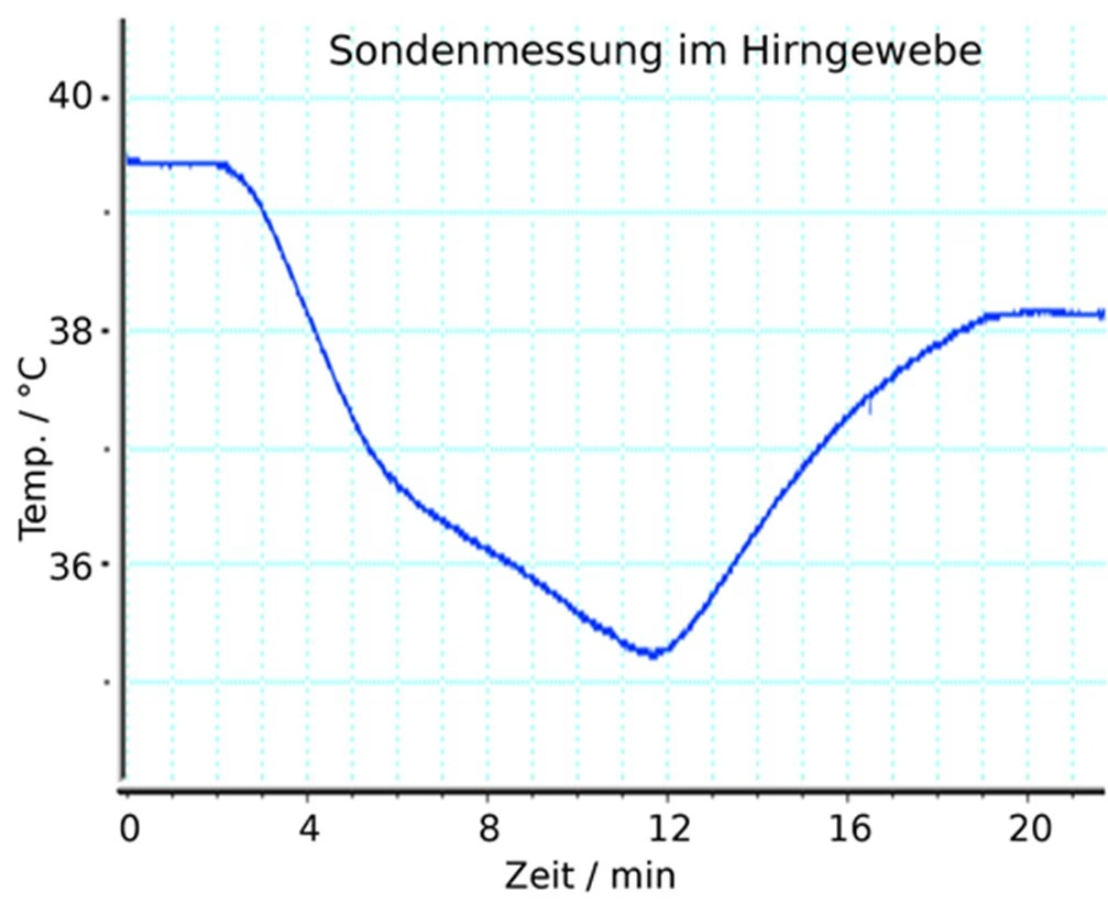
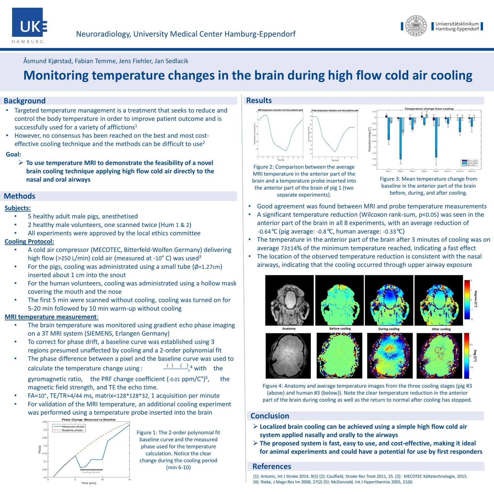
The chart above is a summary. These are the images it was made from — published and unpublished, both shown.
The indication map
Two indications where cooling is standard. Six where it is not.
Cooling is not one therapy with one answer. Timing, thermal distribution, route, population and dose determine what is actually being tested. The map below shows where benefit is established, where signals support further work and which study-design boundaries AIRCHILL must address prospectively.
The clearest positive result in the entire field. Eleven randomised trials in 1,505 term and late-preterm newborns: death or major neurodevelopmental disability at 18 months fell by a quarter — RR 0.75 (95 % CI 0.68–0.83), number needed to treat 7. Cooling must begin within six hours of birth, runs at 33.5 °C for 72 hours, and is a whole-body in-hospital therapy. It does not travel: in low- and middle-income settings the HELIX trial found no benefit and excess mortality, 42 % versus 31 % (p = 0.022).
Jacobs SE, Berg M, Hunt R et al. Cooling for newborns with hypoxic ischaemic encephalopathy. Cochrane Database Syst Rev 2013, CD003311.pub3 · Thayyil S, Pant S, Montaldo P et al. HELIX. Lancet Glob Health 2021;9:e1273–e1285.
Rapid active cooling is standard, and speed is written into the guideline: reach target within 30 minutes of recognition, at a rate of at least 0.155 °C per minute. The certainty behind it is very low, most of the data come from healthy athletes with induced hyperthermia rather than heatstroke patients, and the measured endpoint is cooling rate, not survival. And the goal here is removing excess heat, not inducing hypothermia in a normothermic patient — so it supports a timing argument only by analogy.
Barletta JF, Palmieri TL, Toomey SA et al. SCCM Guidelines for the Treatment of Heat Stroke. Crit Care Med 2025;53:e490–e500 · Douma MJ, Aves T, Allan KS et al. First aid cooling techniques for heat stroke. Resuscitation 2020;148:173–190 · ILCOR CoSTR, first aid cooling techniques, 2019.
Ten randomised trials, 4,220 patients: no change in neurological recovery, RR 1.04 (0.93–1.15), with a harm signal on re-arrest, RR 1.19 (1.00–1.41). The 2025 ERC / ESICM guideline recommends against prehospital cooling by rapid infusion of large volumes of cold fluid after return of circulation. Our own claim register has said the same since the first version, and we build nothing on it.
Lindsay PJ, Buell D, Scales DC. Pre-hospital cooling after out-of-hospital cardiac arrest. Crit Care 2018;22:66 · Kim F, Nichol G, Maynard C et al. JAMA 2014;311:45–52 · ERC-ESICM Post-Resuscitation Care Guidelines 2025.
PRINCESS cooled transnasally during CPR, a median of 19 minutes after collapse, in 677 patients: survival with CPC 1–2 at 90 days 16.6 % versus 13.5 %, difference 3.1 % (95 % CI −2.3 to 8.5), p = 0.25. The cooling worked — time to below 34 °C fell from 182 to 105 minutes — the outcome did not follow. RINSE, cold saline during CPR in 1,198 patients, closed at 48 % of target recruitment and found worse return of circulation in shockable rhythms, 41.2 % versus 50.6 % (p = 0.03).
Nordberg P, Taccone FS, Truhlář A et al. PRINCESS. JAMA 2019;321:1677–1685 · Bernard SA, Smith K, Finn J et al. RINSE. Circulation 2016;134:797–805.
The European Stroke Organisation is explicit: it does not recommend induction of hypothermia to improve functional outcome or survival — weak recommendation, very low quality, resting on six trials and 252 patients. The trial meant to settle it, EuroHYP-1, stopped in 2018 after 98 of a planned 1,500 patients, and only 31 % of cooled patients ever reached the protocol temperature. It failed on feasibility, not on biology. For a device company that is a different finding, and the more interesting one.
Ntaios G, Dziedzic T, Michel P et al. ESO Guidelines for the Management of Temperature in Patients with Acute Ischemic Stroke. Int J Stroke 2015;10:941–949 · van der Worp HB, Macleod MR, Kollmar R et al. EuroHYP-1. Eur Stroke J 2019;4:254–262 · CORDIS final report, EU project 278709.
Thirty-seven randomised trials in 3,110 patients — and Cochrane declined to pool them, because the results point in different directions. GRADE very low. Eurotherm3235 was stopped early because outcomes were worse with cooling (mortality 34.9 % versus 26.6 %). POLAR started cooling a median of 1.8 hours after injury — about as fast as anyone has managed in an adult trial — and found 48.8 % versus 49.1 %. Speed alone did not rescue it. This is an important boundary condition for our thesis: early systemic hypothermia in an unselected severe-TBI population was not beneficial, so any AIRCHILL TBI programme would need a distinct thermal exposure and a prospectively selected phenotype.
Lewis SR, Evans DJW, Butler AR et al. Hypothermia for traumatic brain injury. Cochrane Database Syst Rev 2017, CD001048.pub5 · Andrews PJD, Sinclair HL, Rodriguez A et al. Eurotherm3235. N Engl J Med 2015;373:2403–2412 · Cooper DJ, Nichol AD, Bailey M et al. POLAR. JAMA 2018;320:2211–2220.
One randomised, unblinded pilot: 55 adolescent athletes, two 30-minute head-and-neck cooling sessions at 6 °C, faster symptom recovery on SCAT5 (p = 0.003), no serious adverse events. The authors call it a feasibility study, and so do we. No guideline recommends cooling for concussion.
Congeni J, McCulloch J, Swanson J et al. Head and neck cooling therapy after concussion. Clin J Sport Med 2022;32:341–347.
Active temperature control is recommended; cooling to 33 °C is not established over 36.8 °C. THAPCA-OH: survival with good function at one year 20 % versus 12 %, relative likelihood 1.54 (95 % CI 0.86–2.76), p = 0.14. THAPCA-IH was stopped for futility at 329 of 558 patients: 36 % versus 39 %, RR 0.92 (0.67–1.27).
Moler FW, Silverstein FS, Holubkov R et al. THAPCA-OH. N Engl J Med 2015;372:1898–1908 · THAPCA-IH. N Engl J Med 2017;376:318–329 · ILCOR, post-arrest temperature management in children, 2021.
Two indications on this list are standard of care, and they are the two where cooling starts fastest: newborn encephalopathy within six hours of birth, heatstroke within thirty minutes of recognition. Wherever cooling begins hours after the injury, it has not delivered — and wherever it begins fast, it is standard of care. That is an association across indications, not evidence — and it has an obvious counter-example sitting in the same table: POLAR started within two hours and was flatly negative. So this does not prove that speed is the missing variable. It is why we think the question is worth a device, and why our claim register keeps ultra-early selective cooling at clinical hypothesis and not one step higher.
Every figure above was checked against the cited source on 9 August 2026. Two of the sources carry caveats that belong here rather than in a footnote nobody reads: the ILCOR paediatric statement from 2026 is a draft for public comment and is not cited as a guideline, and the neonatal Cochrane review dates from 2013 and remains the current version. Where a source could only be reached through a society summary rather than the publisher, the number is marked in our internal documentation and not stated here as verbatim.
Preclinical timing evidence · Lyden et al.
Across multiple animal models, earlier cooling and the delivered thermal dose can materially change biological and neurological outcomes. In a murine cardiac-arrest model, intra-arrest cooling outperformed delayed post-ROSC cooling and normothermia; in a porcine prolonged-arrest model, selective intranasal cooling started with CPR improved resuscitation success; in a rat asphyxial-arrest model, combined intra- and post-arrest cooling produced the strongest survival and neurological result; and focal-ischaemia experiments show that delay, depth and duration materially influence neuroprotection. These models do not predict AIRCHILL’s human effect size, but they directly support testing a very early, controlled thermal exposure rather than treating late systemic cooling as the same biological question.
Lyden and colleagues combined a randomized rodent middle-cerebral-artery-occlusion model with primary rodent neurovascular-unit cell models. The study is relevant to the AIRCHILL research hypothesis because it directly tested whether when cooling starts, how deep it goes and how long it lasts change neuroprotection. It does not establish AIRCHILL efficacy or a human treatment target.
The standard rodent stroke model used random allocation and 33 °C cooling. Two hours immediately before or after reperfusion produced smaller infarctions than the four-hour post-reperfusion regimen. The overall one-way ANOVA was significant (F3,113=6.6; p<0.001), but Tukey post-hoc testing did not separate the individual groups. That limitation is part of the finding, not a footnote to hide.
Original experiment: Lyden PD et al., J Cereb Blood Flow Metab. 2019;39:1693–1709. Schematic redrawn by Medical Cooling from the published methods and Figure 1 legend; not a reproduction of the publisher figure.
Neuron OGD model · qualitative redraw
2 h
6–24 h
2 h
6–24 h
Primary rodent neurons underwent two hours of oxygen-glucose deprivation and were then assigned to 33, 35 or 37 °C, with delays of 0–90 minutes and treatment durations of 2, 6 or 24 hours. Temperature, delay and duration were all highly significant. The authors report 33 °C as superior to 35 °C for essentially every delay-duration combination except the 90-minute-delay/2-hour condition. Immediate treatment gave the greatest protection; after a 90-minute delay, longer treatment was required to regain cytoprotection.
Qualitative redraw of the published Figure 3 findings. Cell-culture evidence is preclinical and is not equivalent to an in-vivo animal outcome or a human clinical effect.
Lyden PD, Lamb J, Kothari S, Toossi S, Boitano P, Rajput PS. Differential effects of hypothermia on neurovascular unit determine protective or toxic results: Toward optimized therapeutic hypothermia. Journal of Cerebral Blood Flow & Metabolism. 2019;39(9):1693–1709. First published online 21 November 2018. doi: 10.1177/0271678X18814614 · PMCID: PMC6727141. The publisher page provides a permissions-request route; the visualizations above are original Medical Cooling redraws of reported study relationships rather than copies of the journal artwork.
Our own data
Peer-reviewed mechanism data — and the engineering targets they define.
Carried out at the Department of Diagnostic and Interventional Neuroradiology, University Medical Center Hamburg-Eppendorf, under BMBF Go-Bio grant 031A530. Fabian Temme is a co-author on the publication and on the award-winning poster.

MR thermogram of the porcine brain with cooling off and on. Five anaesthetised pigs, 3 T proton-resonance-frequency thermometry. Whole brain −0.33 ± 0.30 °C after five minutes; anterior brain −0.83 ± 0.51 °C, significant in every experiment. 73 ± 14 % of the maximum effect was reached within three minutes.
Sedlacik J et al., Therapeutic Hypothermia and Temperature Management, doi 10.1089/ther.2017.0031.

Anatomy, before, during and after cooling in a healthy volunteer. Two volunteers, three experiments, hollow mask over mouth and nose. −0.33 °C in the inferior frontal gyrus, p < 0.05 in all three experiments; the three other brain regions showed no significant change.
ISMRM 2016, Singapore, Magna cum laude poster award. Conference abstract, not a patient study.

Porcine lung after six hours of ventilation with air at −20 °C through an endotracheal tube. All entities histomorphologically intact: bronchiole, cartilage, blood vessels, alveoli. No pathological findings in lung or brain.

Contrast-enhanced 2D lung perfusion before and after cooled ventilation. No relevant change. MR perfusion gave the same result, and intraprocedural CT showed no evidence of pulmonary oedema. The animals remained cardiorespiratorily stable throughout.
14 pigs, 11 volunteers
Two animal series — 5 pigs for cooling depth and 9 in a further preclinical series — plus a human study with 11 healthy volunteers who tolerated cold air to −20 °C for up to ten minutes. The published subsets are smaller: 5 pigs in the peer-reviewed mechanism paper and 2 volunteers across 3 experiments in the ISMRM 2016 abstract. Multiple additional scientific conference contributions and posters from the AIRCHILL programme are documented; the complete event bibliography will be added after line-by-line verification of the original records.
73 % within three minutes
Averaged over eight experiments the anterior brain reached 73 ± 14 % of its minimum temperature after three minutes. Time to first measurable effect is the property that matters prehospital.
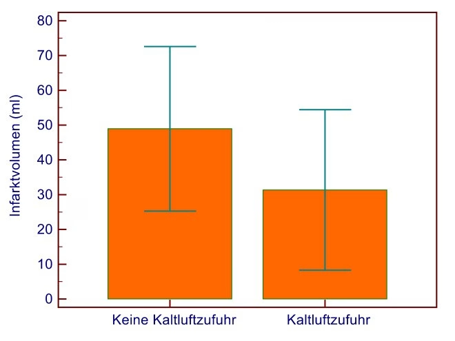
Stroke model
The report notes that infarct development may be limited by immediately initiated cold air therapy. This signal supports prospective replication and helps define an efficacy study; the original experiment was not designed as definitive efficacy proof.
Sedlacik J, Kjørstad Å, Nagy Z, Buhk JH, Behem CR, Trepte CJ, Fiehler J, Temme F. Feasibility Study of a Novel High-Flow Cold Air Cooling Protocol of the Porcine Brain Using MRI Temperature Mapping. Therapeutic Hypothermia and Temperature Management, doi 10.1089/ther.2017.0031 · Final report “Airchill – Gekühlte Beatmung”, BMBF grant 031A530, Department of Diagnostic and Interventional Neuroradiology, University Medical Center Hamburg-Eppendorf, 28 March 2017 · Kjørstad Å, Temme F, Fiehler J, Sedlacik J, ISMRM 24th Annual Meeting, Singapore 2016.
Next engineering objective
Increase and distribute lower-airway heat transfer.
“Eine Kühlung des gesamten Hirns ist mit der evaluierten Methode nicht erreichbar.”
The 2017 tracheal setup was well tolerated and defined the central engineering challenge: its thermal effect was limited by dead-space ventilation, while the endonasal route produced a stronger local effect. That result gives the next programme a measurable objective — increase lower-airway heat transfer while preserving ventilation and airway safety. The endonasal route is thermodynamically what RhinoChill does, and RhinoChill missed its primary endpoint in PRINCESS. The effect we measured is local: our authors attribute it to direct airway exposure rather than cooling of carotid blood, so whole-brain cooling is not established and a comparison with whole-body trials such as TTM2 is not admissible.
We publish this for two reasons. It is the honest reading of our own data, and it defines the engineering problem precisely. Dead-space ventilation is a design constraint, not a law of physics: flow, temperature, timing and circuit geometry were not optimised in 2017. Whether it can be overcome is an open development question — and the reason our evidence plan starts at mechanism rather than outcomes.
An early market entry is planned around a narrow performance and safety claim, not a neurological outcome claim. The latter requires a separate and substantially larger evidence pathway. Sample sizes and claim strategy will be updated after the intended-purpose decision, Notified Body feedback and the gate results.
Research only · therapeutic gas admixtures
Therapeutic-gas biology opens a second research axis for AIRCHILL.
AIRCHILL has no clinical patient data with hydrogen, argon, xenon, helium, nitric oxide, carbon monoxide, nitrous oxide or hydrogen sulfide. No evidence currently demonstrates that combining AIRCHILL respiratory cooling with any therapeutic gas improves survival, neurological function or recovery. The table below separates human signals from preclinical hypotheses.
HYBRID II: 2% H₂ for 18 h after OHCA. Primary 90-day CPC 1–2 endpoint 56% vs 39%, p=0.15; positive secondary survival/mRS signal. Hydrogen-FAST is extending clinical delivery research.Human signal · priority 1
Strong porcine post-cardiac-arrest signals support translation: 70% Ar / 30% O₂ for 4 h improved neurological recovery in small randomized pig studies. The first human phase-II CPAr RCT is designed for 120 unconscious shockable-rhythm OHCA survivors, using 70% Ar / 30% O₂ for 4 h and 48-h neuron-specific enolase as the primary endpoint. The EU record was authorised but still listed recruitment as pending in March 2026; patient-outcome benefit therefore remains unestablished.Strong preclinical · human phase II pending
Randomized human post-arrest research has already crossed the biomarker threshold: xenon added to hypothermia reduced white-matter injury on diffusion-tensor MRI, but the study was not powered to establish a neurological-outcome benefit. Translation remains technically demanding because xenon generally requires high inspired concentrations, recirculating/closed-loop delivery and careful anaesthesia and haemodynamic integration.Randomized human biomarker signal
Established inhaled pulmonary vasodilator with experimental cerebral-perfusion and reperfusion rationale. Small post-cardiac-arrest human feasibility work has shown that inhaled NO can be delivered in this setting, but no randomized evidence establishes improved neurological outcome after cardiac arrest, stroke or TBI. Any AIRCHILL programme would therefore treat NO first as a tightly monitored adjunct requiring patient-near NO/NO₂/FiO₂ measurement and methemoglobin surveillance.Human feasibility · neuro efficacy unproven
Clinical neuroprotection remains unproven, but helium has a uniquely relevant engineering signal for AIRCHILL: human inhalation is feasible and a 2025 porcine respiratory-cooling study reported substantially faster brain cooling when helium replaced nitrogen as the carrier gas. That makes helium especially interesting as a thermal/flow medium whose value should be tested first by heat-transfer, gas-exchange and ventilator-performance endpoints rather than by assuming a pharmacological neuroprotective effect.Human feasible · thermal carrier priority
Mechanistic or preclinical signals exist, but current human neuro-efficacy and safety profiles do not justify a clinical AIRCHILL programme.No-go for clinical programme
A potent physiological cerebral vasodilator, but the 1,700-patient TAME trial did not improve functional neurological outcome. Treat PaCO₂ as a ventilation target, not a new therapeutic-gas claim.Device parameter only
The H₂ case is unusually close to the AIRCHILL architecture: a low 2% inspired concentration has already been delivered through mechanical ventilation after cardiac arrest in randomized human research. But HYBRID II was small and prematurely terminated, and its primary neurological endpoint was not statistically significant. Its secondary survival signal is a reason to test, not a reason to claim efficacy.
A future translational programme should separate cooling alone, gas alone and cooling + gas. Otherwise a positive or negative result cannot tell whether the thermal intervention, the medicinal gas or their interaction caused the finding.
Hydrogen requires dedicated ignition, leak and oxygen-enrichment engineering. Argon, xenon and helium can displace oxygen and alter flow-sensor behaviour. NO requires patient-near NO/NO₂/FiO₂ monitoring and methemoglobin controls. Xenon and helium require a dedicated thrombolysis-interaction gate before any stroke programme. All gases must be verified behind the cooling stage under worst-case temperature, pressure, humidity and condensation conditions.
Key research sources: HYBRID II, Circ J / eClinicalMedicine post-arrest H₂ programme (PMID 36969346); Hydrogen-FAST protocol (PMID 41204320); Xenon after cardiac arrest, JAMA 2016 / PMID 26978207; argon post-arrest translational literature and CPAr; inhaled NO clinical pharmacology and experimental neurovascular studies; TAME, N Engl J Med 2023. None studied AIRCHILL. Research concentrations shown here are descriptions of study protocols, not treatment recommendations.
Claim register
Every claim we make, and exactly how strong it is.
Each line states what we assert and the level of evidence behind it. No statement anywhere in our materials is stronger than its level here. The limitations of our own work are set out in full in the sections above, and the complete internal register — including the claims we have retired — is part of the material we share on request.
Levels taken from the project’s single source of truth, 8 August 2026. Claims about other interventions — routine or late cooling to 33 °C, transnasal cooling during resuscitation — are covered in the clinical-context section above rather than repeated here. External evidence: Kim F et al., JAMA 2014 · Dankiewicz J et al., N Engl J Med 2021 · Nordberg P et al., JAMA 2019 · Taccone FS et al., Crit Care 2021 and Crit Care 2024;28:335 · ERC-ESICM Guidelines 2025 · § 137e SGB V and BT-Drs. 21/6808.
Claim policy
How we state development goals responsibly.
We lead with what has already been measured and with the next study objective. AIRCHILL’s clinical programme is designed to establish patient safety, reproduce product-level thermal performance and test whether the rapid early effect seen in preclinical and exploratory human work translates into patient benefit. Until those studies report, these remain validation objectives rather than established clinical outcomes.
Read the study material yourself.
The publication, the final report and the poster are available on request, together with our assessment of what they do and do not support.